A year ago today, I was lying on an operating table in Indianapolis while a very good surgeon sliced out a lymph node chock full o'teratoma. Time flies when you're having fun! Today, the scar looks OK, although the top part is a bit uglier than the bottom part (my guess would be that several of the urology interns took turns stapling me back together after surgery, and some did a better job than others). It's by far the best $25,000 ever spent on me.
Life goes on. Hallelujah.
Showing posts with label teratoma. Show all posts
Showing posts with label teratoma. Show all posts
Wednesday, May 31, 2006
Wednesday, May 24, 2006
Coming soon on DVD!
[Fade in...]
Pathologist: "It's cancer."
[Dissolve to...]
Pathologist: "Sorry, I was smoking crack, is isn't cancer."
[Cut to title with voiceover]
Announcer: "Who cares what the hell it is?"
Check it out on video (it's in four parts, this is part one of four). No, it's not my actual surgery, but a close facsimile thereof. Although not for the squeamish, I happen to find it very cool.
Anyhoo, yesterday was the follow-up with the thoracic surgeon. Very uneventful, although after looking at the chest x-ray that was taken right before my appointment, that chunk of lung he took out seemed a lot larger than I had previously visualized (even though it was probably only about 2% of my total lung tissue).
A nurse took my BP and said, "119 over 72. That's awesome!" I smiled smugly at The Rev., who proceeded to roll her eyes so far back into her head that I did in fact see her optic nerves.
Awesome? Well, yes, but I already knew that.
Oh, in answer to the question I had the other day, the doc said they got the chunk out by deploying some sort of bag with a drawstring around the specimen, then cinching it tight before taking it out through the thorascopy port. (They had deflated my lung in order to operate on it, using a breathing tube and a ventilator to keep my right lung working. The lung tissue folds easily when deflated.)
Part four of the video shows how this happens. The diseased area is isolated with staples and removed from the rest of the lung, then placed in a bag to avoid contamination of other areas of the chest cavity. Cool!
At the end of the day, I was sent home and told the surgeon didn't need to see me again. I felt like I had been spurned following a one-night stand. It was the same story I had heard from my urologist as well as the surgeon who did the RPLND. Why are these surgeons so afraid of commitment?
Back to surveillance!
Pathologist: "It's cancer."
[Dissolve to...]
Pathologist: "Sorry, I was smoking crack, is isn't cancer."
[Cut to title with voiceover]
Announcer: "Who cares what the hell it is?"
It's
Frank's Thoracoscopy!
Check it out on video (it's in four parts, this is part one of four). No, it's not my actual surgery, but a close facsimile thereof. Although not for the squeamish, I happen to find it very cool.
Anyhoo, yesterday was the follow-up with the thoracic surgeon. Very uneventful, although after looking at the chest x-ray that was taken right before my appointment, that chunk of lung he took out seemed a lot larger than I had previously visualized (even though it was probably only about 2% of my total lung tissue).
A nurse took my BP and said, "119 over 72. That's awesome!" I smiled smugly at The Rev., who proceeded to roll her eyes so far back into her head that I did in fact see her optic nerves.
Awesome? Well, yes, but I already knew that.
Oh, in answer to the question I had the other day, the doc said they got the chunk out by deploying some sort of bag with a drawstring around the specimen, then cinching it tight before taking it out through the thorascopy port. (They had deflated my lung in order to operate on it, using a breathing tube and a ventilator to keep my right lung working. The lung tissue folds easily when deflated.)
Part four of the video shows how this happens. The diseased area is isolated with staples and removed from the rest of the lung, then placed in a bag to avoid contamination of other areas of the chest cavity. Cool!
At the end of the day, I was sent home and told the surgeon didn't need to see me again. I felt like I had been spurned following a one-night stand. It was the same story I had heard from my urologist as well as the surgeon who did the RPLND. Why are these surgeons so afraid of commitment?
Back to surveillance!
Friday, May 19, 2006
More fun in CT land
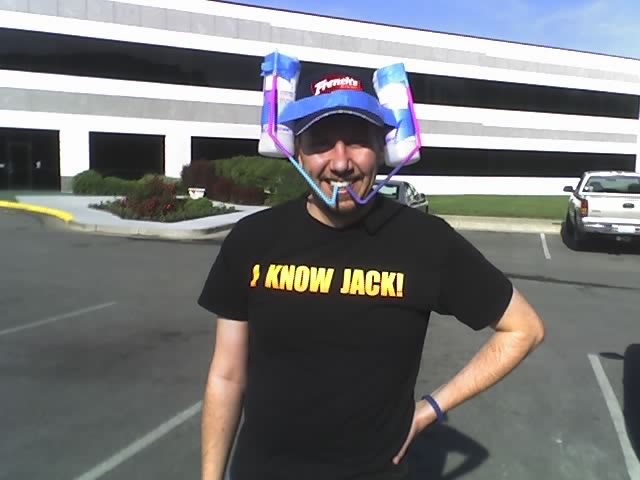 I went back to the Cancer Box for my first post-lung surgery CT scan today. They had me pre-medicate this time because of the hive that continually shows up on my forehead during CTs after the iodine contrast is injected, so I was well-doped up on Benadryl when I came in. Guess what - it didn't help a damned bit, the hive showed up again, and all I got was a sleepless night to add to my cranky morning since I couldn't eat beforehand. Still, I managed to make a new hat for the occasion...
Those are CT contrast bottles taped to my hat, by the way - the smoothie that tastes like artifically flavored berries mixed with dirt in a creamy white base. The proper medical term is "barium sulfate suspension". According to Wikipedia, other uses for barium sulfate include paint pigment (it replaced white lead) and pyrotechnics. Just thought you might like to know. I suppose it could be worse - sometimes the barium sulfate is given by enema rather than orally (usually for colon and small intestine studies).
Anyway, the CT looks good - you can now see staples where the mass used to be. We also got the pathology opinion from IU; they agreed with the local folks that the mass was teratoma and not active cancer. Hot dog! We're back on the surveillance schedule. Like Dr. V. said, it was just a speed bump.
Later in Dr. V.'s exam room, fabulous nurse G. scored me some breakfast from a buffet that had been set up for employees. Eggs and sausage - SWEET! Plus, nurse R. from the chemo room came by and got to satisfy her picking jones by pulling out some suture parts from one of the thoracoscopy ports on my left side (the sutures were popping out and keeping the wound from healing completely). Apparently the nurses all have their own wound fetishes. Nurse G. didn't like the picking at all, but apparently she totally goes wild for pus shooting out of an abcess. OK, that was probably TMI, but welcome to my world.
Afterward, I asked for copies of all my paperwork from this year so far so I can start building my next FAA file. They farmed it out to a woman from the medical records room, who came out with the copies and said something like, "Wow, I actually get to meet Mr. Vinny!" Apparently my hats have made me some sort of legend (in my own mind at least).
OK. It's now time for a nap. Tuesday we meet with the thoracic surgeon so he can look at me and say, "Damn, I do good work!" What I want to know is this: the wedge they pulled out was about 6 x 5 x 2.5 cm (the mass inside was only 1.2 x 1 x 0.9 cm). The ports (i.e., cuts) for the scope are only about 3 cm. How'd they get that chunk out?
Have a great weekend!
I went back to the Cancer Box for my first post-lung surgery CT scan today. They had me pre-medicate this time because of the hive that continually shows up on my forehead during CTs after the iodine contrast is injected, so I was well-doped up on Benadryl when I came in. Guess what - it didn't help a damned bit, the hive showed up again, and all I got was a sleepless night to add to my cranky morning since I couldn't eat beforehand. Still, I managed to make a new hat for the occasion...
Those are CT contrast bottles taped to my hat, by the way - the smoothie that tastes like artifically flavored berries mixed with dirt in a creamy white base. The proper medical term is "barium sulfate suspension". According to Wikipedia, other uses for barium sulfate include paint pigment (it replaced white lead) and pyrotechnics. Just thought you might like to know. I suppose it could be worse - sometimes the barium sulfate is given by enema rather than orally (usually for colon and small intestine studies).
Anyway, the CT looks good - you can now see staples where the mass used to be. We also got the pathology opinion from IU; they agreed with the local folks that the mass was teratoma and not active cancer. Hot dog! We're back on the surveillance schedule. Like Dr. V. said, it was just a speed bump.
Later in Dr. V.'s exam room, fabulous nurse G. scored me some breakfast from a buffet that had been set up for employees. Eggs and sausage - SWEET! Plus, nurse R. from the chemo room came by and got to satisfy her picking jones by pulling out some suture parts from one of the thoracoscopy ports on my left side (the sutures were popping out and keeping the wound from healing completely). Apparently the nurses all have their own wound fetishes. Nurse G. didn't like the picking at all, but apparently she totally goes wild for pus shooting out of an abcess. OK, that was probably TMI, but welcome to my world.
Afterward, I asked for copies of all my paperwork from this year so far so I can start building my next FAA file. They farmed it out to a woman from the medical records room, who came out with the copies and said something like, "Wow, I actually get to meet Mr. Vinny!" Apparently my hats have made me some sort of legend (in my own mind at least).
OK. It's now time for a nap. Tuesday we meet with the thoracic surgeon so he can look at me and say, "Damn, I do good work!" What I want to know is this: the wedge they pulled out was about 6 x 5 x 2.5 cm (the mass inside was only 1.2 x 1 x 0.9 cm). The ports (i.e., cuts) for the scope are only about 3 cm. How'd they get that chunk out?
Have a great weekend!
Tuesday, May 02, 2006
The FFR is here!
Got a call from Dr. V. (the onc) today.
me: "What's up?"
Dr. V.: "Well, the 'FFR' is here."
me: "What's that?"
Dr. V.: "The final effin' report."
That's a verbatim quote by the way; I'm not sure the f-word is in Dr. V.'s vocabulary. Anyway, the final pathology results are in from the local lab. Turns out the mass that was removed was not active germ cell cancer after all as the preliminary report said, but was metastasized teratoma, as we had originally expected. Phew! Good news for now.
He had also spoken with the pros from Indianapolis, and they want the pathology slides so they can do their own study and report. Works for me. More news as it develops...
Thursday, January 13, 2005
the low down diagnosis
Here's what we found out on Wednesday Jan 12, 2005 (from Frank's email to Schmink)
the nitty gritty:
the nitty gritty:
--
The pathology report says the tumor was mixed germ cell, 95% embryonal,
less than 5% mature teratoma, focal seminona.
It was classed pT2 (tumor extends through tunica albuginea with vascular/lymphatic invasion).

CT scan shows: "There is a large inhomogeneous mass seen in the left periaortic area that
measures 6 cm x 5 cm in size.  This displaces the left renal vein anteriorly and extends over
routine 7 mm images anterior to the left psoas into the region of the left common iliac artery."
(the orchicetomy was on the left side)  N.B - the onc thinks the size is underestimated by
at least 2 cm (in the head-to-toe measurement).

Bloodwork on 12/27 (10 days before the orchiectomy):
LDH 258, AFP 9897, B-HCG 372

Bloodwork on 1/10 (5 days after the orchiectomy):
LDH 216, AFP 9464, B-HCG 451

So it's non-seminoma that has metastatized into the retroperitoneal lymph system,
with a big frickin mass sitting on my aorta.
The urologist suggests chemo first and maybe surgery later after the mass has shrunk.
The oncologist/hematologist suggests the same, except he will probably insist on
surgery after chemo.

The urologist isn't big into staging; the oncologist said pT2, N3, S2; or stage IIC.

Here's the plan: I go get a port-a-cath next week, then start chemo on the 24th.
3 rounds of BEP (maybe a fourth if the tumor markers don't fall like they should) with Neupogen
between rounds; 12 weeks total (more if 4 rounds).
Intermediate CT scans, with the likelihood of surgery after the chemo is done.
Subscribe to:
Posts (Atom)